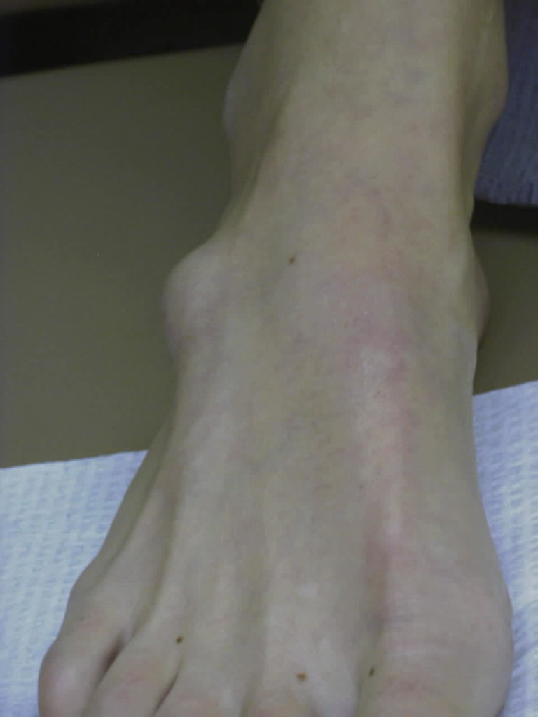
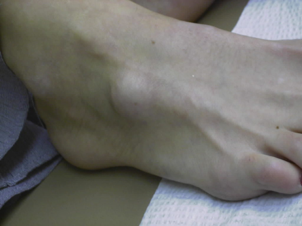
The delightful bump you see on the side of this lady's foot is a ganglion cyst. If you puncture it (and I did) you would see that it is filled with a thick gel. The gel is called synovial fluid better known as joint fluid. It is the material inside every joint and the outside sheath of many tendons that helps to lubricate and nourish the joint. The fluid is held inside the joint by a covering called a capsule. If something causes the capsule to tear just a little the inside bag of fluid can pop out and form a ganglion. Early on ganglions typically get larger some days and smaller other days depending on the activity of the joint. After several months they stay pretty much the same size and start to get harder as the fluid inside gets thicker.
Ganglions can occur anywhere on the body. The ones on the wrist are sometimes called Bible Cysts because of an old fashioned treatment of whacking them with a heavy object, like a book. This thump sometimes ruptured the cyst on the inside and it would go away.
In a more civilized method of doing the same thing we sometimes "incise" them in the office. Using a local anesthetic block on the skin a needle or blade is used to puncture the cyst and the fluid can be expressed out. Replacing the gel with a small quantity of a steroid medication can sometimes get the thing to stay away. Ganglions can be subjected to this "incision and drainage" several times. If it does not go away you might want to have it surgically removed.
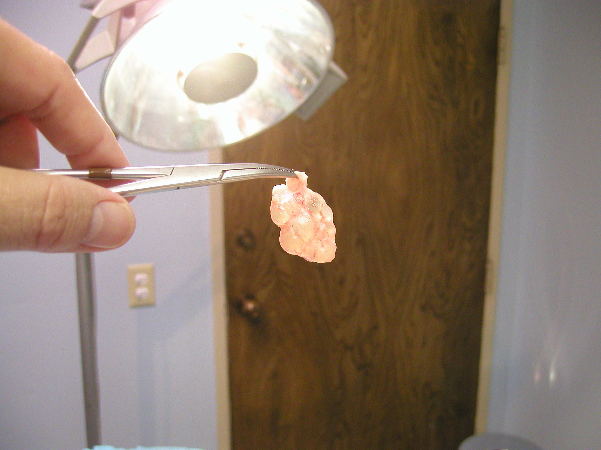
Here is a photo of a large ganglion that was removed from a foot

Ganglions are not visible on plain x-rays but they are sometimes taken to see if there is any bone abnormality under the lesion. Ganglions can be seen with ultrasound examination and with MRI (magnetic resonance imaging). I sometimes ask for a pre-surgical MRI for particularly large ganglions to see if the lesion tracks down to a bone joint or to see if it is arising from a tendon.
Small ganglions can be removed in the office but larger ones, like this one, are done at the surgery center or the hospital using a bit of gentle sedation and local anesthesia (general anesthesia is not needed). The sutures left in the skin after surgical removal are removed in 2 weeks. Most foot surgeons will want you to keep the wound dry for the 2 weeks that the sutures are in the foot. During this time you will have dressings on your foot and will probably be walking in a surgical shoe.
Mucoid cysts are similar and can be found on the toes (and fingers for that matter) by the nails. For pictures and a discussion of Mucoid Cysts please CLICK HERE

